


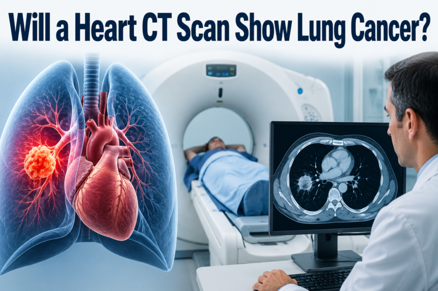
A heart CT scan is not built to check for lung cancer, but since the lungs sit right next to the heart, small parts of lung tissue often appear in the images anyway. Because of this, a heart CT can sometimes pick up an unusual spot or shadow in the lung purely by accident.
However, this scan is never meant to replace a proper lung cancer screening, since its settings, focus, and image quality are all tuned for the heart and not the lungs. If you truly want to know your lung health status, a dedicated chest CT or low-dose CT is the correct choice, and this article explains exactly why.
What Is a Heart CT Scan (Cardiac CT)?
A heart CT scan, also called a cardiac CT, is a specialized imaging test used to closely examine the heart and its blood vessels. It is mainly used to detect blockages, plaque buildup, and calcium deposits that could lead to heart disease or a heart attack.
Purpose of a Cardiac CT
The main goal of a cardiac CT is to give doctors a clear picture of how blood is flowing through the coronary arteries. It helps identify narrowed or blocked vessels that could increase the risk of chest pain or cardiac events, allowing doctors to plan treatment early instead of waiting for symptoms to worsen.
Types of Heart CT Scans
There are two commonly used versions of this scan, and each serves a slightly different purpose depending on what the doctor is trying to evaluate in the patient’s heart health.
| Scan Type | Main Use |
| Coronary CT Angiography (CCTA) | Detects blockages and narrowing in coronary arteries using contrast dye |
| Calcium Score CT | Measures calcium buildup in arteries without using contrast dye |
What This Scan Is Designed to Detect
A cardiac CT is designed strictly around heart-related structures such as arteries, valves, and calcium deposits. It is not calibrated to study lung tissue in detail, which is one of the biggest reasons people ask, will a heart CT scan show lung cancer, since the scan simply isn’t built with that goal in mind.
What a Heart CT Scan Actually Captures?
Even though a heart CT scan focuses on cardiac structures, the scanning machine still passes through the chest area, meaning parts of the lungs unavoidably show up in the images. This is simply due to anatomy, not because the scan is designed to study lungs.
Understanding the Scan’s Field of View
The “field of view” refers to the total area captured during imaging. Since the heart sits in the middle of the chest, surrounded by lung tissue on both sides, some lung sections naturally appear at the edges of the scan, even though they are not the primary target.
Why Parts of the Lungs Appear in the Images
Doctors do not intentionally include lung tissue in a cardiac CT, but because the heart and lungs are physically close together, complete exclusion is not possible. What appears is often only a partial or lower-quality view rather than a full detailed scan of the lungs.
Why Lung Detail Isn’t the Main Focus
The scanning protocol for a heart CT uses different slice thickness and contrast timing compared to a lung-focused scan. These technical settings are optimized to highlight blood vessels and calcium, not soft lung tissue, which means small nodules can easily be missed or appear unclear.
Can a Heart CT Scan Detect Lung Cancer?
A heart CT scan can occasionally reveal a visible lung nodule or mass by chance, but this is considered an incidental finding rather than a confirmed diagnosis. It should never be treated as a reliable method for detecting or ruling out lung cancer.
Incidental Nodules and Masses
Sometimes, radiologists notice a spot on the lung tissue captured during a cardiac scan, even though they were not specifically looking for it. This is known as an incidental finding, and while it can be an early warning sign, it usually requires further testing to confirm what it actually is.
Why Incidental Findings Are Not the Same as Screening
Finding something by chance is very different from actively screening for a disease. A screening test is designed with settings and precision specifically meant to catch early lung changes, while an incidental finding is simply a byproduct of a scan meant for something else entirely.
Real-World Frequency of Incidental Lung Findings
Studies on cardiac CT scans have shown that incidental lung nodules are discovered in a noticeable percentage of patients undergoing heart imaging. However, most of these nodules turn out to be harmless, and only a small portion require closer monitoring or additional testing.
Why a Heart CT Isn’t a Substitute for Lung Cancer Screening
Relying on a heart CT scan alone for lung cancer detection can create a false sense of security, since the scan simply isn’t built to catch early or small lung changes with accuracy.
Differences in Scan Resolution and Settings
A dedicated lung screening scan uses thinner image slices and settings specifically adjusted to highlight lung tissue clearly. A heart CT, on the other hand, prioritizes vascular structures, which means finer details in the lungs may not be visible at all.
Small Nodules Can Be Missed Easily
Because the cardiac scan protocol isn’t tuned for lung visibility, very small or early-stage nodules can go completely unnoticed. This is one of the biggest risks of assuming a heart scan can double as a lung check.
The Danger of False Reassurance
Many patients wrongly believe that a “clear” heart CT scan also means their lungs are healthy. This false reassurance can delay proper lung screening, allowing a potential issue to grow silently in the background.
What Scan Actually Detects Lung Cancer?
To properly check for lung cancer, doctors recommend scans that are specifically designed around lung tissue rather than the heart or other organs.
Low-Dose CT (LDCT) for Lung Cancer Screening
A low-dose CT scan is currently the gold standard for lung cancer screening, especially for people at higher risk. It uses a reduced amount of radiation while still capturing highly detailed images of lung structures, making it both safe and effective for regular screening.
Diagnostic Chest CT
A diagnostic chest CT is used when a patient already has symptoms or a suspicious finding that needs closer investigation. Unlike LDCT, this scan usually involves more detailed imaging and sometimes contrast dye to get a clearer picture of any abnormality.
Who Should Get Screened
Screening guidelines generally recommend regular lung checks for older adults with a significant smoking history or other major risk factors. A doctor can help determine individual eligibility based on age, health history, and lifestyle factors.
What Happens If Something Is Found on a Heart CT?
If a lung nodule or unusual spot is noticed during a heart CT, doctors typically recommend a proper follow-up rather than ignoring it, since early evaluation can make a significant difference in outcomes.
Next Steps After an Incidental Nodule
The doctor may suggest a follow-up chest CT after a few months to see if the nodule has changed in size or shape. In some cases, additional tests like a biopsy may be recommended if the nodule appears suspicious.
Referral to a Specialist
Depending on the nodule’s characteristics, patients are often referred to a pulmonologist or oncologist for a more detailed evaluation. This ensures that any potential issue is handled by a specialist familiar with lung-related conditions.
Why Ignoring Incidental Findings Is Risky
Even a small or seemingly harmless nodule should not be brushed aside without proper follow-up. Ignoring such findings can allow a potentially serious condition to progress unnoticed until it becomes harder to treat.
Heart CT vs. Chest CT vs. Low-Dose CT — Comparison Table
Understanding the difference between these three scans makes it much easier to know which one is right for a specific health concern.
| Feature | Heart CT | Diagnostic Chest CT | Low-Dose CT (Screening) |
| Main Purpose | Heart and artery evaluation | Detailed lung investigation | Early lung cancer screening |
| Radiation Dose | Moderate | Higher | Low |
| Resolution Focus | Cardiac vessels | Full lung detail | Lung nodules and tissue |
| Best For | Heart disease risk | Symptomatic patients | High-risk, routine screening |
Common Myths and Misconceptions
Many misunderstandings exist around what a heart CT scan can and cannot detect, and clearing these up can help patients make smarter healthcare decisions.
“If My Heart Scan Was Clear, My Lungs Are Fine”
This is one of the most common misconceptions, but a clear heart scan only confirms cardiac health, not lung health, since the two scans focus on entirely different structures and settings.
“Any CT Scan Can Detect Cancer”
While CT scans in general are powerful imaging tools, each type is optimized for a specific purpose. A scan not designed for lung tissue will not reliably catch early cancer changes, regardless of how advanced the machine is.
“I Don’t Need a Separate Lung Scan If I’ve Had a Heart Scan”
This assumption can be dangerous, especially for high-risk individuals like long-term smokers. A separate, dedicated lung screening remains essential regardless of any previous heart imaging results.
When Should You Ask for a Dedicated Lung Cancer Screening?
Certain risk factors make it especially important to ask a doctor about lung cancer screening rather than relying on incidental findings from unrelated scans.
Key Risk Factors to Consider
People with a long smoking history, older age, family history of lung cancer, or exposure to harmful substances at work should proactively discuss screening options with their healthcare provider.
Talking to Your Doctor
Bringing up personal risk factors during a routine checkup can help doctors decide whether a low-dose CT screening is appropriate. Being proactive about this conversation can lead to much earlier detection if any issue exists.
FAQs
Can a CT scan for the heart miss lung cancer? Yes, since the scan settings are not designed for lung tissue, small or early-stage nodules can easily be missed during a cardiac CT.
What’s the difference between a cardiac CT and a chest CT? A cardiac CT focuses on heart structures and arteries, while a chest CT is specifically designed to capture detailed images of lung tissue.
How often should high-risk individuals get lung cancer screening? Most guidelines recommend yearly low-dose CT screening for high-risk individuals, though a doctor should confirm the right frequency based on personal health history.
Does a heart CT scan use contrast, and does that affect lung visibility? Some heart CT scans do use contrast dye, but it is timed specifically for blood vessels, which does not necessarily improve lung tissue visibility.
Is a lung nodule found on a heart CT usually cancerous? Most nodules found incidentally turn out to be harmless, but follow-up testing is still necessary to confirm what the nodule actually is.
Conclusion
So, will a heart CT scan show lung cancer? The honest answer is that it might occasionally catch something by chance, but it was never designed for that purpose.
For accurate and reliable lung cancer detection, a dedicated chest CT or low-dose CT screening remains the proper choice.
Anyone with risk factors should speak directly with their doctor rather than depending on incidental findings from an unrelated scan.
Related Posts
Can Being Sick Cause Acne? Causes, Symptoms, and Complete Skin Guide
Can Varicose Veins Cause Knee Pain? Causes, Symptoms & Relief
How Long Does Cataract Surgery Take? Complete Timeline & Recovery Guide
How Long After Fluoride Treatment Can I Eat?
When Do Pregnancy Cravings Start? Complete Timeline & Causes Guide


