


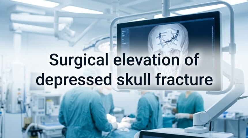
Surgical elevation of depressed skull fracture is a neurosurgical procedure performed to lift a broken piece of skull bone that has been pushed inward below the normal skull surface.
Doctors usually recommend this surgery when the bone depression is deeper than the thickness of the skull, when it presses on the brain, or when it causes a tear in the protective brain lining. The main goal of this surgery is to relieve pressure on the brain, prevent infection, and restore the natural shape of the skull.
Not every depressed fracture needs surgery, and many minor cases heal on their own with monitoring. However, when neurological symptoms, open wounds, or brain compression are present, surgical elevation becomes necessary to avoid long-term complications. This guide explains the entire process in simple, easy-to-understand language.
What Is a Depressed Skull Fracture?
A depressed skull fracture happens when a broken piece of bone shifts inward instead of staying level with the rest of the skull. Below, we explain the different types and common causes behind this injury.
Types of Skull Fractures
Skull fractures are generally divided into closed and open types. In a closed fracture, the skin above the break remains intact, so there is a lower risk of infection reaching the brain. In an open or compound fracture, the skin is torn, exposing the bone and sometimes the brain lining to outside bacteria, which raises the urgency for treatment. The table below highlights the difference clearly.
| Fracture Type | Skin Condition | Infection Risk | Typical Urgency |
| Closed Depressed Fracture | Intact skin | Lower | May allow monitoring |
| Open/Compound Depressed Fracture | Torn or broken skin | Higher | Usually urgent surgery |
Causes and Risk Factors
Most depressed skull fractures result from strong, direct impacts to the head. Common causes include car or motorcycle accidents, falls from height, sports injuries, and physical assaults involving blunt objects. Children and elderly individuals often face higher risk due to thinner bone structure or increased chances of falling, making awareness of head safety especially important for these groups.
Signs and Symptoms
Recognizing the signs early can make a major difference in treatment outcomes. The symptoms usually fall into two categories: neurological and physical.
Neurological Symptoms
Neurological symptoms can indicate that the brain itself is affected by the injury. These may include confusion, sudden weakness on one side of the body, seizures, and difficulty speaking clearly. In more severe cases, loss of consciousness or unequal pupil size may occur, both of which require emergency medical attention right away.
Physical/Visible Signs
Physically, a depressed fracture may show as a visible dent or indentation on the scalp, sometimes accompanied by swelling, bruising, or an open wound. Clear fluid leaking from the nose or ears can indicate a cerebrospinal fluid leak, which is a serious sign that should never be ignored and needs immediate evaluation.
Diagnosis Process
Doctors rely on imaging and physical evaluation to determine how serious the fracture is and whether surgery is required.
Imaging Techniques
Different imaging methods help doctors see the exact location and depth of the fracture. A CT scan is the most commonly used tool because it clearly shows bone displacement and any bleeding inside the skull. X-rays may be used for a quick initial check, while an MRI is sometimes added when soft tissue or brain injury needs closer examination. The comparison table below shows how these methods differ.
| Imaging Method | Best Used For | Speed | Detail Level |
| CT Scan | Bone fractures & bleeding | Fast | High |
| X-ray | Initial quick screening | Very fast | Low to moderate |
| MRI | Soft tissue & brain detail | Slower | Very high |
Severity Assessment Criteria
Doctors typically consider a fracture significant enough for surgery when the depression is deeper than the thickness of the skull bone itself, often cited as more than 5 to 10 millimeters. Other deciding factors include whether the dura, the protective membrane around the brain, has been torn, and whether the wound is contaminated with dirt or debris.
When Is Surgery Needed?
Not every depressed fracture requires an operation. The decision depends on specific medical indicators that doctors assess carefully.
Indications for Surgical Elevation
Surgery is generally recommended when the bone depression is deep enough to press on brain tissue, when there is a confirmed dural tear, or when the patient shows neurological symptoms like seizures or weakness. Open fractures with a high risk of infection almost always require prompt surgical treatment as well.
When Non-Surgical Management Is Appropriate
In cases where the depression is minimal, there is no dural involvement, and the patient shows no neurological symptoms, doctors may choose careful observation instead of immediate surgery. Regular monitoring with follow-up scans helps ensure the injury is healing properly without complications.
The Surgical Elevation Procedure
Understanding what happens during surgery can help ease anxiety for patients and family members preparing for the operation.
Pre-Surgical Preparation
Before surgery, the medical team reviews imaging scans to plan the exact approach and confirm the fracture’s location. The patient is given general anesthesia, and the surgical area is cleaned and prepared to reduce infection risk during the procedure.
Step-by-Step Surgical Process
The surgeon creates an opening around the fractured area, known as a craniotomy, to access the depressed bone safely. The bone fragments are then carefully lifted back into their normal position. If the dura is torn, it is repaired at this stage, and any blood clots or debris underneath are cleaned out before closing the site.
Bone Fragment Management
If the bone piece is healthy and undamaged, surgeons often reposition it and secure it using small plates and screws. However, if the bone is too damaged or contaminated to reuse safely, it may be removed and replaced later through a separate procedure called cranioplasty, using synthetic material or a bone graft.
Recovery and Post-Surgical Care
Recovery time and care needs vary depending on the severity of the original injury and how the surgery was performed.
Immediate Post-Op Care
Right after surgery, patients are closely monitored in a hospital setting, often in an intensive care unit, to watch for signs of swelling, bleeding, or seizures. Pain management and infection prevention through antibiotics are also standard parts of early recovery.
Long-Term Recovery Timeline
Full recovery can take anywhere from several weeks to a few months, depending on the injury’s severity and the patient’s overall health. Some patients may need physical or cognitive rehabilitation if the brain was affected, while others recover with minimal lasting effects.
Risks and Complications
Like any brain surgery, this procedure carries certain risks that patients should understand beforehand.
Surgical Risks
During and shortly after surgery, risks include infection at the incision site, excessive bleeding, and cerebrospinal fluid leakage. Surgeons take strict precautions to minimize these risks, but they remain an important part of informed consent discussions.
Long-Term Complications
Over time, some patients may experience seizures, changes in skull shape, or in rare cases, cognitive difficulties related to the original brain injury rather than the surgery itself. Regular follow-up appointments help catch and manage these issues early.
Cost Considerations
The cost of surgical elevation for a depressed skull fracture varies widely based on hospital type, geographic location, severity of the injury, and length of hospital stay.
Emergency surgeries, ICU care, and any additional procedures like cranioplasty can significantly affect the overall cost, so discussing payment options and insurance coverage with the hospital beforehand is strongly advised.
Frequently Asked Questions
Is surgery always required for a depressed skull fracture?
No, surgery is only needed when the depression is significant, when brain compression exists, or when the dura is torn.
How long does recovery take after this surgery?
Recovery can range from a few weeks to several months, depending on the injury’s severity and the patient’s overall health condition.
What imaging test is best for diagnosing this condition?
A CT scan is typically the fastest and most reliable method for identifying bone displacement and internal bleeding.
Can the original bone always be reused during surgery?
Not always. If the bone is too damaged or contaminated, doctors may remove it and replace it later using cranioplasty.
What are the warning signs that need emergency care?
Confusion, seizures, unequal pupils, and clear fluid leaking from the nose or ears all require immediate medical attention.
Does this surgery guarantee full recovery?
Not necessarily. Outcomes depend on how severe the original brain injury was, along with how quickly treatment was received.
Are children more at risk for depressed skull fractures?
Yes, children often have thinner skull bones, which can make them more vulnerable to this type of injury from falls or accidents.
Conclusion
Surgical elevation of a depressed skull fracture plays a critical role in protecting the brain from long-term damage when the injury is severe enough to require intervention.
Early diagnosis through imaging, combined with timely surgical care, greatly improves recovery outcomes.
Anyone showing signs of a skull injury, especially with neurological symptoms, should seek emergency medical evaluation immediately rather than waiting for symptoms to worsen.
Related posts
How to Restore Gum Health Naturally and Professionally?
Spectra S2 Breast Pump: The Complete Guide for New and Pumping Moms
Are Allergies Hereditary? Understanding Genetics, Risk Factors & Evidence-Based Prevention
How to Fix Peeling Skin on Feet: Causes, Remedies & Treatment
Does Invisalign Hurt? A Complete Guide to Pain, Discomfort, and Relief Tips


